

Summary
“Myxoid changes are the leading cause of mitral valve (MV) prolapse and regurgitation,” according to Brian Griffin, MD, Cleveland Clinic. Myxoid chordae in MR have <25% of normal load bearing capacity, Dr. Griffin noted. “But are these myxomatous changes genetically programmed or maladaptative?” Dr. Griffin asked. “More studies are needed to help answer this question”
- valvular
“Myxoid changes are the leading cause of mitral valve (MV) prolapse and regurgitation,” according to Brian Griffin, MD, Cleveland Clinic. Myxoid chordae in MR have <25% of normal load bearing capacity, Dr. Griffin noted. “But are these myxomatous changes genetically programmed or maladaptative?” Dr. Griffin asked. “More studies are needed to help answer this question”
Timing of Surgery
Timely—but appropriate—surgical intervention has become the key to better outcomes. Raphael Rosenhek, MD, Ludwig Boltzmann Institute for Cardiovascular Research, Vienna, Austria, asked: “When should we operate in MR?” Considerations include pre-op determinants of post-op outcomes, and the likelihood of success in repair vs. replacement, Dr. Rosenhek noted.
A key pre-op factor is ejection fraction (EF). “EF >60% bodes well for operative success,” said Dr. Rosenhek. “But EF <50% is not so encouraging.”
Dr. Rosenhek recommended surgery when symptoms appear, or in asymptomatic patients with LV dilation or falling EFs. “Otherwise, watchful waiting is acceptable.”
A complementary presentation by Robert O. Bonow, MD, Chief, Division of Cardiology, Northwestern University Medical School, said that U.S. guidelines were essentially similar. “There is a paucity of data regarding MR, so expert consensus is the principal basis for guidelines.”
The key is operating early enough to preclude deteriorating symptoms and early death, observed Dr. Bonow, “but late enough to justify risks of surgical intervention.” In Dr. Bonow's view, the natural history of MR now favors early intervention. “One must continue to weigh the anticipated outcome of surgery against the expected outcome of either watchful waiting or medical management alone.”
Pecutaneous MV Repair
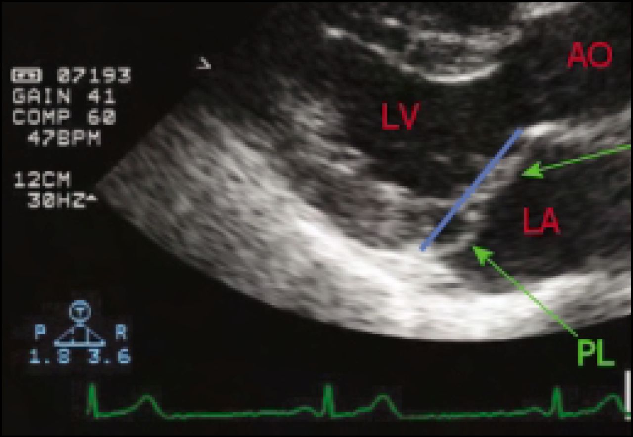
Allan Schwartz, MD, Chief, Division of Cardiology, Columbia University, reviewed surgical alternatives. “The Alfieri approach was first used about 10 years ago,” he said. “This is an edge-to-edge method that brings together the middle scallops of the mitral leaflets to create a double orifice.”
The technique has gained popularity over the last decade, and has extended indications for mitral valve repair.
“But percutaneous approaches are gaining ground and may ultimately prove the best option for repair in many patients,” said Dr. Schwartz.
Dr. Schwartz cited EVEREST II, which he termed “a pivotal study of percutanous techniques in MV surgery.” EVEREST II evaluated a percutaneous method to create the same type of repair as the Alfieri method using the “mitra-clip.”
Dr. Schwartz shared vivid animated imagery demonstrating the mitra-clip's insertion and placement in the left atrium. The clip is then centered over the valve orifice, passed just beyond into the left ventricle and pulled back to grasp the mitral leaflets. After verification that MR is reduced, the clip is released.
In EVEREST II clips were placed in 24 patients. In 13 of 14 patients with reduction of MR to </= 2+ at 30 days post-clip, the clip repair was intact at 6 months.
EVEREST II demonstrated that percutaneous edge-to-edge mitral valve repair can be performed safely. “If you can get out to 3 months post-clip without problems, the odds are good it will stick,” said Dr. Schwartz.
- © 2006 MD Conference Express
Tools


Table of contents
Cited By...
- No citing articles found.