

Summary
Limited range of hip internal rotation has been associated with an increased risk for anterior cruciate ligament (ACL) injury, whereby the hips of ACL-injured patients rotated 12.6° less than those of noninjured individuals [Gomes JL et al. Arthroscopy 2008]. This article discusses the underlying mechanism of injury to the ACL associated with decreased internal rotation of the hip.
- Hip & Knee Conditions
- Sports Medicine Clinical Trials
- Orthopaedics
- Hip & Knee Conditions
- Sports Medicine
- Orthopaedics Clinical Trials
Mélanie Beaulieu, MSc, a doctoral student at the University of Michigan School of Kinesiology, Ann Arbor, Michigan, USA, reported on the underlying mechanism of injury to the anterior cruciate ligament (ACL) associated with decreased internal rotation of the hip.
Limited range of hip internal rotation has been associated with an increased risk for ACL injury, whereby the hips of ACL-injured patients rotated 12.6° less than those of noninjured individuals [Gomes JL et al. Arthroscopy 2008]. The most common cause is femoroacetabular impingement, a condition in which 1 or both bones of the hip joint are abnormally shaped. This deformity can produce abnormal contact between the bones, which progressively damages the joint. The prevalence of femoroacetabular impingement is 6% to 25% in individuals who are asymptomatic [Monazzam S et al. Bone Joint J 2013; Reichenbach S et al. Arthr Care Res 2010] but exceeds 60% in patients with pathological hips [Beck M et al. J Bone Joint Surg Br 2005]. Affected individuals are predominantly young (eg, college athletes) and have an increased risk for early osteoarthritis of the hip.
The study was grounded in 2 hypotheses. The first posited that as internal femoral rotation decreases, the magnitude of peak ACL strain increases. The second was that women have greater peak ACL strain than do men, regardless of the range of internal femoral rotation.
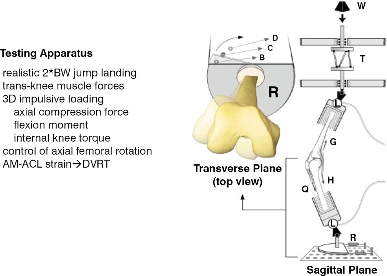
Twenty knee specimens, 10 each from men and women, were harvested from fresh, unembalmed cadavers and tested. The donors were similar in age (men, 59.9 ± 6.6 years; women, 55.2 ± 10.5 years), with men being predictably taller (men, 1.77 ± .05 m; women, 1.67 ± .06 m), heavier (men, 81.3 ± 8.2 kg; women 60.5 ± 8.3 kg), and with greater body mass indexes (men, 25.9 ± 2.3 kg/m2; women 21.6 ± 3.1 kg/m2) than women. Each knee was dissected to leave the joint capsule and associated tendons of the quadriceps, hamstrings, and gastrocnemii intact. Each knee specimen was inverted and positioned in a testing apparatus, diagrammed in Figure 1. The device was designed to subject the specimen to forces that simulate a jump landing at twice body weight, including a twist of the knee (ie, internal tibial torque). The forces, moments, and motion produced during the landings were measured. Also, ACL strain was measured using a device called a differential variable reluctance displacement transducer, which was placed on the anteromedial bundle of the ACL. For each knee specimen, 4 conditions of internal femoral rotation were simulated, ranging from locked to free rotation (∼3°–15°).

Diagram of the Testing Apparatus
AM-ACL=anteromedial anterior cruciate ligament; BW=body weight; DVRT=differential variable reluctance transducer.
Reproduced with permission from M Beaulieu, MSc.
As expected, peak ACL strain increased as internal femoral rotation was decreased during the simulated pivot landings. Furthermore, the female ACLs experienced greater peak strain than did the male ACLs, irrespective of the range of internal femoral rotation.
The researchers surmised that the cause of the increased ACL strain is the increased internal rotation and anterior translation of the knee joint that occur. The authors postulated that screening for a limited range of hip internal rotation might be helpful in identifying athletes with increased risk for ACL injury. These athletes may benefit most from participating in ACL injury prevention programs.
- © 2014 MD Conference Express®
Tools


Table of contents
Cited By...
- No citing articles found.