

Summary
Myositis is an autoimmune disease affecting skeletal muscle, skin, heart, and lungs. The autoantibody directed against histidyl tRNA synthetase (Jo1) is specific for myositis and is the most common autoantibody found in these patients. This article discusses research in the characterization of Jo1-specific T helper cell responses in blood and bronchoalveolar lavage samples obtained from patients with myositis.
- Inflammatory Disorders
- Rheumatology Clinical Trials
- Rheumatology
- Inflammatory Disorders
- Rheumatology Clinical Trials
Myositis is an autoimmune disease affecting skeletal muscle, skin, heart, and lungs. Types of myositis include polymyositis, dermatomyositis, inclusion body myositis, and immune mediated necrotizing myopathy. This is a rare disorder, occurring with an incidence of approximately 1 in 100,000/year. While typically treated with steroids and other immuno-suppressive drugs, the search continues for effective therapies. The autoantibody directed against histidyl tRNA synthetase (Jo1) is specific for myositis and is the most common autoantibody found in these patients. Approximately 20% [Gunawardena H et al. Rheumatology (Oxford) 2009] of myositis patients will have detectable Jo1 autoantibodies and its presence is associated with interstitial lung disease (ILD). Evidence suggests that the autoimmune response of myositis originates in the lungs [Chinoy H et al. Ann Rheum Dis 2012].
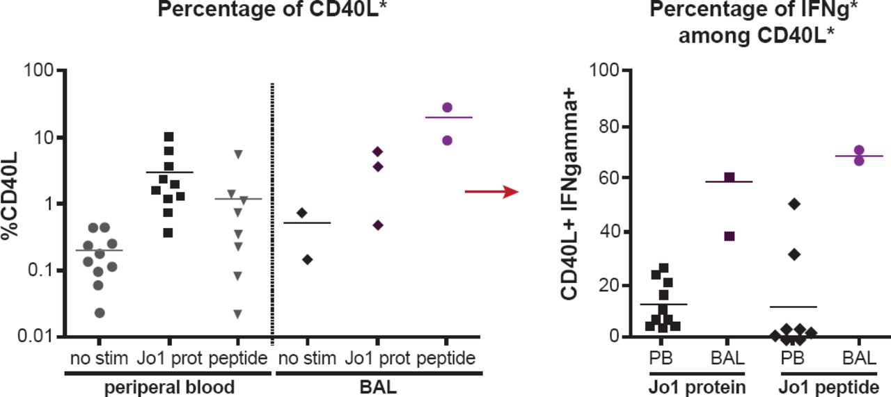
Inka Albrecht, PhD, Karolinska University Hospital, Stockholm, Sweden, presented her research in the characterization of Jo1-specific T helper cell responses in blood and bronchoalveolar lavage (BAL) samples obtained from patients with myositis. Building on earlier published research that revealed the existence of a Jo1-antigen T cell response [Ascherman DP et al. J Immunol 2002], Prof. Albrecht and colleagues sought to determine the frequency and phenotype of the cells, their location, and the major T cell epitope. A 13 amino-acid peptide was derived from the first 60 amino acids of the full Jo1 protein. Cells isolated from the peripheral blood mononuclear cells (PBMCs; n=9) and BAL (n=3) of patients with myositis were stimulated with the Jo1 protein and the test peptide [Chattopadhyay PK et al. Nat Med 2005; Frentsch M et al. Nat Med 2005]. CD154, also known as CD40 ligand (CD40L), is a marker that is upregulated on the cell when the antigen-specific Th cell recognizes its antigen. The expression of the pro-inflammatory cytokines interferon (IFN)-γ, interleukin (IL)-2, and IL-17A were also analyzed. Cells from PBMCs stimulated with the full Jo1 protein had increases in CD40L, IFN-γ, IL-2, and IL-17A compared with unstimulated cells. Similar results were obtained when the cells were stimulated with the test peptide. Blocking assays determined that this reaction was specific. When the PBMC results from all tested patients were pooled, the CD40L increases were significant when stimulated with the full protein (p<0.0001) but not with the test peptide (p=0.1307; Figure 1). This suggests other epitopes that lie outside of the test peptide sequence may be involved. BAL cells also had increased CD04L, IFN-γ, IL-2, and IL-17A when stimulated with the full Jo1 protein and the test peptide, with dramatically higher increases in IFN-γ compared with PBMCs.

Summary of Flow Cytometry Results
Reproduce with permission from I Albrecht, PhD.
Further characterization revealed that the BAL Th cells are Th1 with high expression of chemokine receptors CCR5 and CXCR3. “In the lung the cells have a clear pro-inflammatory phenotype and we were able to identify the first major epitope within the first 60 amino acids,” summarized Prof. Albrecht.
Future research will focus on analyzing cells from the blood and BAL of additional Jo1-positive patients, establishing a tetramer for the identified epitope, and searching for additional epitopes. It is hoped that further identification, isolation, and characterization of Jo1-specific CD4 T cells will ultimately lead to novel therapies for myositis.
- © 2013 MD Conference Express®
Tools


Table of contents
Cited By...
- No citing articles found.