

Summary
Myocarditis is a broad diagnosis based on the presence of tissue inflammation. Making a pathologic diagnosis is difficult with the nonspecific methods available. This article discusses the benefits of cardiac MRI for diagnosing myocarditis.
- Imaging Modalities
- Cardiac Imaging Techniques
- Inflammatory Disease
- Magnetic Resonance Imaging
Myocarditis is a broad diagnosis based on the presence of tissue inflammation. Making a pathologic diagnosis is difficult with the nonspecific methods available. Daniel M. Couri, MD, Southern Illinois University School of Medicine, Springfield, Illinois, USA, discussed the benefits of cardiac MRI (cMRI) for diagnosing myocarditis.
Cardiac troponin T (cTnT) can be elevated in more than 30 conditions. Assomull et al. [Eur Heart J 2007] recruited 60 consecutive patients within 3 months of presentation with ST elevation, normal coronary arteries, and cTnT elevation. After cMRI, an identifiable basis for troponin elevation was established in 65% of patients. The most common underlying cause was myocarditis (50%). Cardiac MRI has broad performance advantages with regard to morphology, function, and the unique ability to directly visualize tissue characteristics [Freidrich MG et al. J Am Coll Cardiol Img 2008].
In the acute phase of myocarditis (Days 1 to 7), the virus spreads to the heart with minimal myocyte damage and necrosis. The subacute phase (up to 30 days) is characterized by viral shedding, cellular damage, and the secondary immune response with antibody generation, cell destruction, inflammation, and hyperemia. Advanced fibrosis predominates in chronic myocarditis, with interstitial replacement, myocardial dilation, and heart failure.
Tissue characterization with cMRI is primarily based on T2- and T1-weighted imaging. T2 imaging focuses on hyperemia and edema in the acute and subacute phases. A diffuse pattern or patchy hyperenhancement scattered throughout the myocardium is consistent with myocarditis. T2 imaging can be quantitated by indexing it to the skeletal muscle; an increase in the edema ratio (ER) ≥2 times that in skeletal muscle is significant.
T1-weighted cardiac imaging is based on gadolinium (Gd) enhancement, which shortens the T1 time and accentuates T1 tissue characteristics. Gd is an extracellular agent with low infiltration into normal myocardium, where myocytes are densely packed and there is little extracellular space. Gd has a high myocyte infiltration rate in damaged myocardium and significantly prolonged infiltration in fibrotic areas. T1 imaging shows early enhancement in the acute and subacute phases, revealing cell damage, hyperemia, and extracellular matrix changes. Late enhancement is focused on focal fibrosis and remodeling.
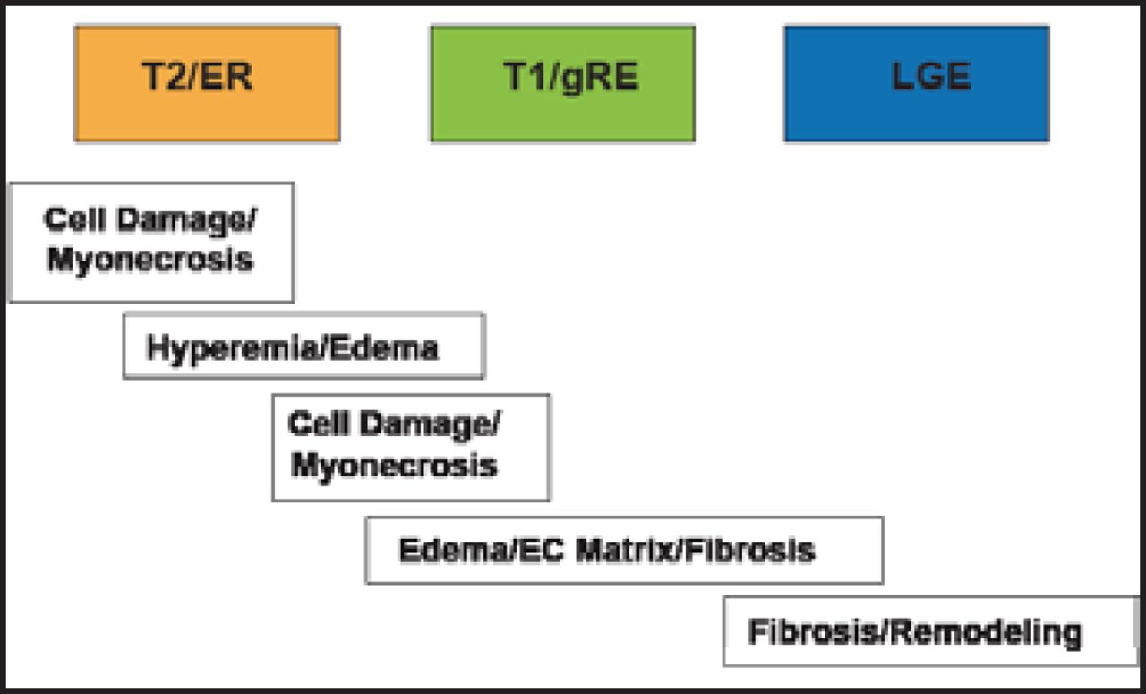
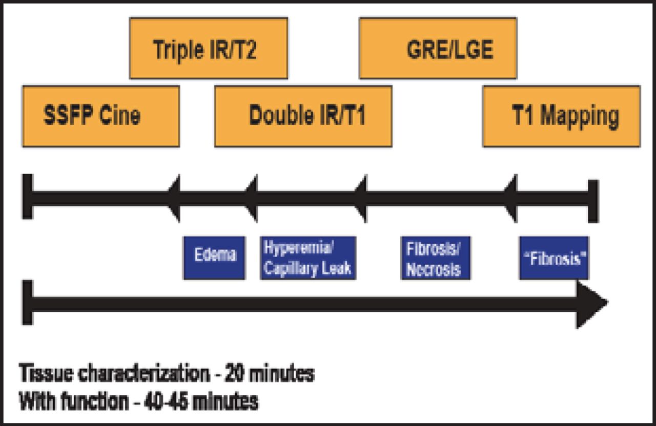
A comprehensive cMRI protocol for diagnosing and assessing myocarditis involves T2 imaging with ER in the acute and subacute phases, T1 imaging with early Gd enhancement ratio (gRE) in the subacute phase, and late Gd enhancement (LGE) in the chronic phase (Figure 1). Figure 2 shows the sequence of cMRI techniques, with steadystate free-precession (SSFP) cine, triple inversion recovery (IR)/T2 (edema), double IR/T1 (hyperemia/capillary leak), GRE/LGE (fibrosis/necrosis), and T1 mapping (fibrosis).

Comprehensive cMRI Protocol for Myocarditis.
EC=endothelial cell; ER=edema ratio; gRE=Gd; enhancement ratio; LGE=late Gd enhancement.

Comprehensive cMRI Protocol Sequence.
IR=inversion recovery; SSFP=steadystate free-precession.
The Lake Louise Consensus Criteria require at least 2 of the following: relative enhancement index (T2) ≥2.0, increased global myocardial gRE ≥4.0, at least 1 focal LGE lesion with nonischemic regional distribution [Friedrich MG et al. J Am Coll Cardiol 2009]. These criteria were developed from pooled data from <300 patients, with no multicenter data. Sensitivity is 69%, specificity is 91%, and accuracy is 78%. Table 1 shows the diagnostic performance of global myocardial edema, global relative myocardial enhancement, LGE, and Lake Louise Criteria.
Diagnostic Performance of Global Myocardial Edema, Global Relative Myocardial Enhancement, Late Gd Enhancement, and Lake Louise Criteria.
Dr. Couri concluded that noninvasive imaging offers an efficient and safe means for acute management of patients. Cardiac MRI is the most versatile and powerful imaging modality for the comprehensive assessment of cardiac pathology. Despite its current success, however, cMRI in myocarditis remains a work in progress.
- © 2012 MD Conference Express®
Tools


Table of contents
Cited By...
- No citing articles found.