

Summary
The integration of imaging modalities may provide a broader picture of disease anatomy, burden, and molecular activity. This article discusss the utility of magnetic resonance and computed tomography imaging in the setting of electrophysiology.
- tomography
- cardiac imaging techniques
- magnetic resonance imaging
- imaging modalities
The integration of imaging modalities may provide a broader picture of disease anatomy, burden, and molecular activity. Edward T. Martin, MD, Oklahoma Heart Institute, Tulsa, OK, discussed the utility of magnetic resonance (MR) and computed tomography (CT) imaging in the setting of electrophysiology.
One of the main factors in deciding whether a patient is a candidate for an implantable cardioverter defibrillator is whether the left ventricular ejection fraction is below 35%. Accurate ejection fraction assessment can be difficult for a 2-D technique, like echocardiography, especially in patients with prior myocardial infarction. A 3-D imaging modality, such as MRI, offers a more accurate method to quantify left ventricular function, which can save lives as well as money.
Using either MR or CT imaging prior to radiofrequency ablation for atrial fibrillation allows the clinician to evaluate the anatomy and assess for the presence of a common ostium and the number, position, and location of pulmonary veins, reducing the risk of complications. Additionally, these CT or MR images may be incorporated into the fluoroscopic images for a highly detailed intraprocedure evaluation, which may also reduce radiation exposure and fluoroscopic time. In a study by Ector and colleagues, CT images of the pulmonary veins, merged with fluoroscopic images, resulted in detailed anatomical and electrical activation maps [Ector J et al. Circulation 2005; Ector J et al. Circulation 2007]. Delayed enhancement MRI of the left atrium prior to a radiofrequency ablation procedure for atrial fibrillation can predict postprocedure atrial fibrillation recurrence, noted Dr. Martin [Oakes RS et al. Circulation 2009].
Zahi Fayad, PhD, FAHA, FACC, Translational and Molecular Imaging Institute, Mt. Sinai School of Medicine, New York, NY, discussed the benefit of multimodality imaging, particularly with regard to MRI and fluorodeoxyglucose positron emission tomography (FDG-PET). The benefit of this combination imaging technique is that it is highly reproducible and noninvasive and provides quantitative data about vessel morphology, metabolic activity and glycolysis, tissue composition, and inflammation. The level of radiation that is used with CT is decreasing, but the exposure can be further reduced with combination MRI/PET imaging, Dr. Fayad noted. However, the protocol for this combination is more complex and may require more experience and/or close attention to the guidelines for this technique.
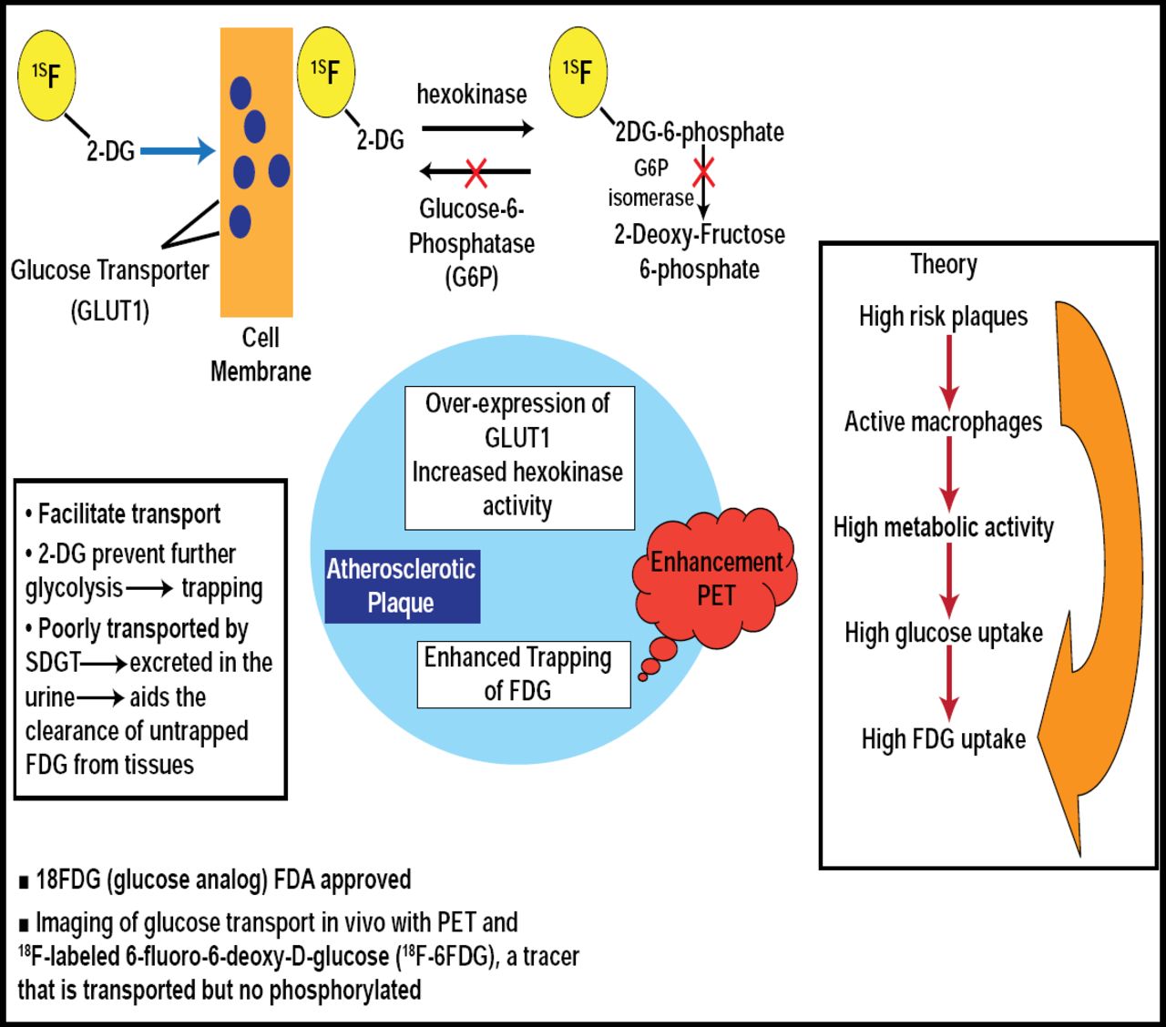
Studies have shown that FDG-PET is a useful tool for detecting molecular markers of inflammation and plaque vulnerability, which may have implications for stroke prevention and carotid atherosclerotic intervention assessment (Figure 1) [Tawakol A et al. J Am Coll Cardiol 2006; Graebe M et al. Eur J Vasc Endovasc Surg 2009]. Strong correlations have been found with FDG-PET uptake in carotid plaques and gene expression of the macrophagespecific marker CD68 (p=0.02). Weaker correlations have been found with cathepsin K, matrix metalloproteinase 9, and interleukin 18 gene expression.

PET/CT Detection of Plaque.
The use of statins in atherosclerosis is not uncommon. In addition to the FDG-PET gene expression correlation, this technique, coupled with MRI, may be useful in monitoring anti-inflammatory effects of statins over time [Fayad ZA et al. J Am Coll Cardiol 2005; Tahara N et al. J Am Coll Cardiol 2006]. Using MRI and FDG-PET as evaluation tools, Dr. Fayad is currently assessing the efficacy of a drug on atherosclerotic plaque in an ongoing multicenter Phase II trial, the results of which are still pending.
In December 2009, the merging of imaging techniques was realized. The first whole-body multimodality scanner was introduced at the Mt. Sinai Medical School of Medicine. This technology allows CT, PET, and MRI to occur in the same setting. Although the cost of such technology may be an issue at the moment, Dr. Fayad explained that the benefit is significant, and as with many new technologies, the cost often decreases over time, especially once it enters mainstream clinical medicine.
- © 2010 MD Conference Express
Tools


Table of contents
Cited By...
- No citing articles found.