

Summary
Treatment with KIOM-79, a novel mixture of 4 herbal extracts, appears to slow the development of retinopathy, according to a new animal study of diabetic eye disease. In addition to characterizing the treatment effects of KIOM-79, the study provides important insights into the pathophysiology of diabetic retinopathy.
- retinal diseases
- diabetes mellitus
- endocrinology
Treatment with KIOM-79, a novel mixture of 4 herbal extracts, appears to slow the development of retinopathy, according to a new animal study of diabetic eye disease. In addition to characterizing the treatment effects of KIOM-79, the study provides important insights into the pathophysiology of diabetic retinopathy.
Vascular Damage in Retinopathy
Researchers have previously observed the accumulation of advanced glycation end products (AGEs) in the neural retina and vascular cells of diabetic animals (Miura J et al. J Diabetes Complications 2003). In these animals, AGE accumulation appears to induce the programmed death of retinal pericytes and neuronal cells. Retinal pericyte apoptosis leads to a range of damaging events within the retina, including the development of microaneurysms, retinal hemorrhages, neovascularization, and permanent impairment of visual function.
KIOM-79 contains the extracts of 4 herbs: parched Puerariae radix and gingered Magnoliae cortex, Glycyrrhizae radix, and Euphorbiae radix. These agents inhibit AGE-induced apoptosis by preventing NFkB activation and lowering proapoptotic cytokine production. Jin Sook Kim, MD, Korea Institute of Oriental Medicine, Daejeon, Republic of Korea, described the effects of KIOM-79 on AGE-induced retinal damage.
Treatment Effects of KIOM-79
In the current study, Dr. Kim and colleagues treated 7-week-old male Zucker Diabetic Fatty (ZDF) rats with KIOM-79 (50 mg/kg) or placebo once daily for 13 weeks. Normal, untreated rats also were included as control animals. At the end of the treatment period, the retinas were harvested and examined for signs of vascular damage.
Compared with rats in the vehicle-treated group, KIOM-79-treated rats had significantly lower serum levels of AGEs (p<0.05) and lower numbers of AGE-positive retinal cells (p<0.05).
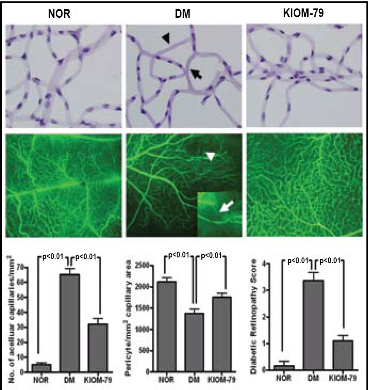
Retinas from the vehicle-treated group showed evidence of vascular damage when assessed by immunohistochemistry, including areas of retinal pericyte loss and the appearance of acellular capillaries. However, these vascular changes rarely were observed among retinas that were harvested from KIOM-79-treated rats (Figure 1, top panel).
Additional fluorescein staining allowed researchers to calculate retinal angiography lesion scores, which are an indication of the degree of retinopathy. When assessed by fluorescein staining, retinas from vehicle-treated ZDF rats showed severe signs of vascular damage, including vessel-narrowing, fluorescein leakage, and nonperfusion of fluorescein. By contrast, retinas from KIOM-79-treated diabetic rats showed significantly fewer changes in retinal angiography (p<0.05; Figure 1, middle panel).
These findings provide quantitative evidence to support the further study of KIOM-79, Dr. Kim said. “Treatment with KIOM-79 is useful in inhibiting the accumulation of AGEs in retinal tissue and has a preventive effect on the development of diabetic retinopathy,” she concluded.

Effects of KIOM-79 on Retinal Vascular Damage.
Top panel: Immunohistochemistry of retinal samples showing pericyte ghost (arrow) and acellular capillary (arrowhead).
Middle panel: Fluorescein angiography showing nonperfusion areas and vessel-narrowing (magnified inset).
Bottom panel: Quantitative analysis of acellular formation, pericyte ghosts, and retinopathy score of retinal vessels. Nor = normal, untreated rat; DM = ZDF rat treated with placebo; KIOM-79 = ZDF rat treated with KIOM-79.
- © 2008 MD Conference Express
Tools


Table of contents
Cited By...
- No citing articles found.