

Summary
Jeroen J. Bax, MD, 2009 ESC Congress Programme Committee Chair, University Hospital Leiden, Leiden, The Netherlands, discusses new trends and advancements in diagnostic cardiac imaging
- imaging modalities
- cardiac imaging techniques
The field of diagnostic cardiac imaging has experienced many exciting innovations and advances in recent years. Each modality has clinical advantages and disadvantages and their use in clinical practice varies greatly. The main imaging modalities include echocardiography, magnetic resonance imaging (MRI), nuclear cardiology and multi-slice computed tomography (MSCT).
Echocardiography is the most commonly used imaging technique in clinical cardiology due to its reliability and practicality. This technique is highly reproducible, mobile and available bedside. It provides an accurate evaluation of cardiac dimensions and function, valvular anatomy and function, cardiac pressures, and the pericardium without the use of radiation.
Stress echocardiography (physiological exercise or pharmacological stress) is used for detection of myocardial ischemia as a marker of coronary artery disease (CAD). Intravenous contrast can be used to enhance left ventricular border opacification, as well as to evaluate myocardial perfusion.
Magnetic resonance imaging (MRI) produces information similar to echocardiography but with a higher resolution. With contrast-enhanced imaging, high precision imaging of scar tissue is possible without radiation. The most significant drawbacks to this technique are claustrophobia, the presence of cardiac pacemakers and defibrillators, and limited availability of equipment.
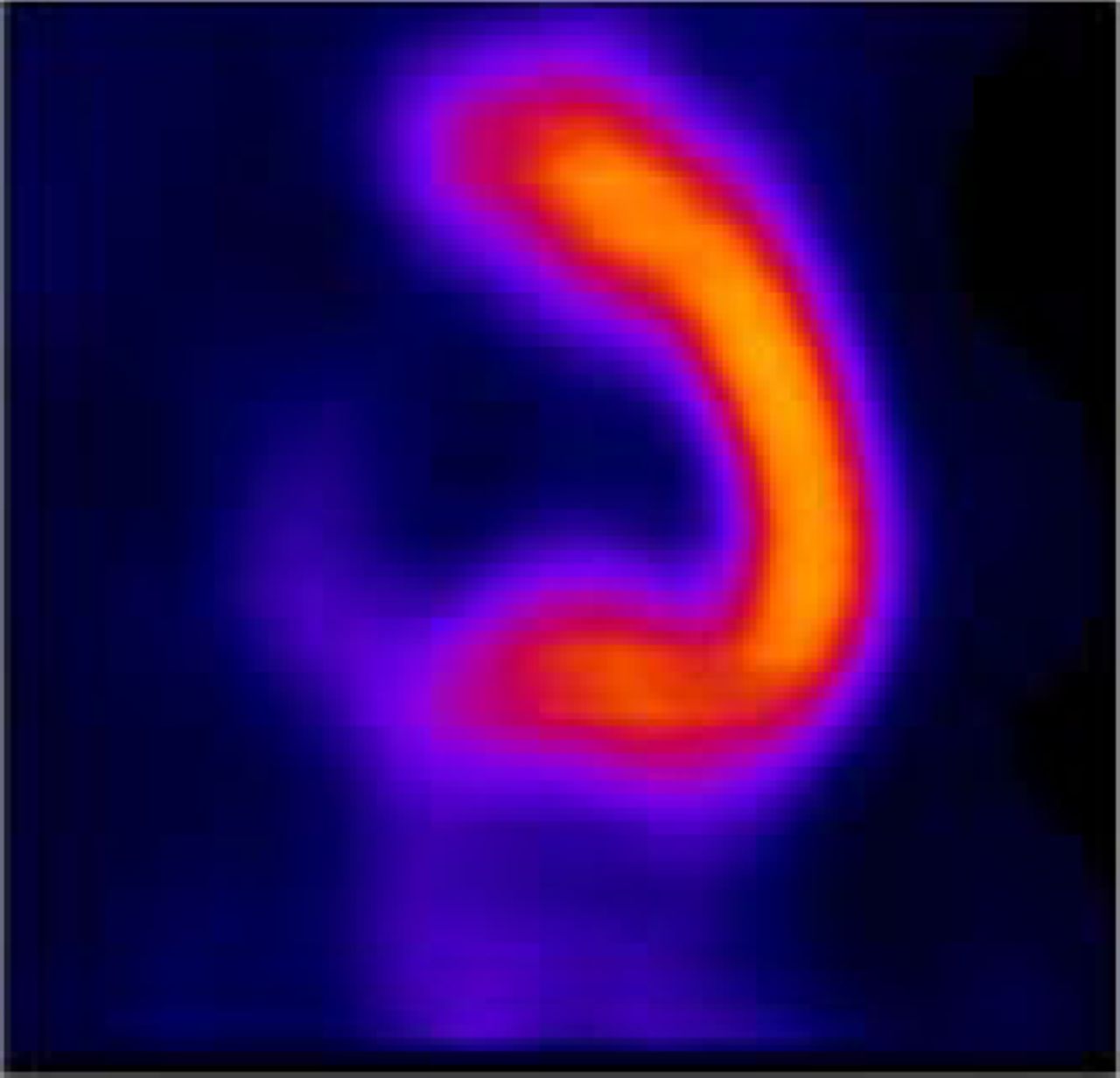
Nuclear cardiology includes imaging with PET and SPECT, the two most frequently used techniques for assessment of myocardial ischemia. Radioactive tracers are used to visualize cardiac biology and functional processes. The main focus of SPECT imaging is assessment of myocardial perfusion (Figure 1A); in addition, assessment of cardiac innervation is also possible. The main advantage of PET imaging is the option of absolute quantification of physiological processes such as perfusion, metabolism and innervation.

SPECT Perfusion Study.
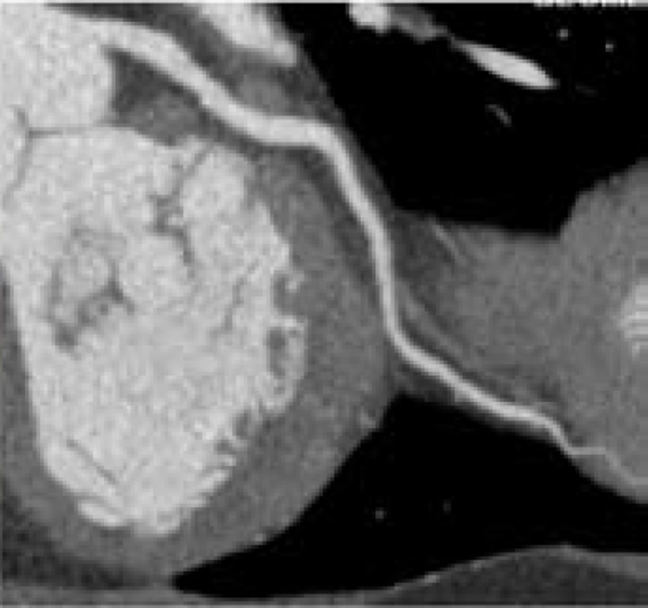
The most recent advance in imaging is MSCT. This technique provides assessment of coronary atherosclerosis (a calcium score) and permits non-invasive angiography (Figure 1B). It provides anatomic imaging, without information on the hemodyamic consequences of the observed coronary lesions (ie, no information on the presence/absence of ischemia secondary to the detected atherosclerosis is provided). This technique utilizes contrast agents and radiation. The main advantage is that CAD can be assessed at a very early stage (ie, atherosclerosis without significant coronary artery stenoses). It is clear that MSCT has a high diagnostic accuracy for detection/exclusion of CAD, but its precise role in clinical evaluation and patient work-up remains to be determined.

MSCT Angiography.
Accordingly, the current era is characterized by a large variety of non-invasive imaging techniques, and the (near) future will determine the precise roles of these various imaging techniques.
The upcoming Annual Meeting of the European Society of Cardiology (Aug 30 -Sept 3, 2008, Munich, Germany; http://escardio.org/congresses), will emphasize clinical cardiology and imaging. Presentations including “Expert Imagers” and Live-Case Imaging sessions will specifically address imaging technology and how it can be integrated to support and enhance clinical care. One of the most exciting new features being introduced this year is the “Everything You Wanted to Know About Cardiovascular Imaging but were Afraid to Ask” session. This new interactive session will allow attendees to post questions on the ESC website prior to the meeting which will then be answered by our expert panel during the live session, providing a wonderful opportunity for attendees to interact with our experts. I look forward to seeing you in Munich this August!
- © 2008 MD Conference Express
Tools


Table of contents
Cited By...
- No citing articles found.