

Summary
Systemic lupus erythematosus (SLE) and systemic sclerosis (SSc; scleroderma) are autoimmune disorders that belong to a largergroup of connective tissue diseases that can occur at all ages. SLE (lupus) is a chronic autoimmune disorder that can affect virtually any organ of the body. This article discusses the histopathological criteria for renal lupus and the implications for therapy, neuropsychiatric SLE, the relationship between scleroderma and pulmonary arterial hypertension, as well as future treatments, among other topics.
- rheumatological autoimmune disorders
- lupus
- systemic connective tissue disorders

Systemic lupus erythematosus (SLE) and systemic sclerosis (SSc; scleroderma) are autoimmune disorders that belong to a larger group of connective tissue diseases that can occur at all ages. SLE (lupus) is a chronic autoimmune disorder that can affect virtually any organ of the body. It is more common in young women, predominantly during the reproductive age. In patients with SLE, the immune system becomes hyperactive, forming antibodies that attack normal tissues and organs, including the skin, joints, kidneys, brain, heart, lungs, and blood. Although effective therapies exist, the etiology of SLE is unknown, SLE is associated with considerable morbidity and mortality including the development of renal complications. Dr. Franco Ferrario from the S. Carlo Borromeo Hospital in Milan, Italy, reported on the revised histopathological criteria for renal lupus and the implications for therapy (Kidney Int 2004;65:521–30). He stated that the purpose of the revisions was to allow for a clear and unequivocal description of the lesions and classes of lupus nephritis. This improved standardization should support treatment and further clinicopathologic studies.
The new classifications were developed by a group of renal pathologists, nephrologists and rheumatologists. Six classes were proposed with Class I and II being used purely for mesangial involvement. Class III and IV describe focal glomerulonephritis (involving <50% of total number of glomeruli) and diffuse glomerulonephritis (involving ≥50% of total number of glomeruli). Class V is reserved for membranous lupus nephritis, and Class VI for advanced sclerosing lesions. The group further recommended that diagnosis include concomitant vascular or tubulointerstitial lesions.
Neuropsychiatric SLE: An Elusive Syndrome
Dr. Melanie Harrison, Hospital for Special Surgery, NY, discussed the need for better definition of neuropsychiatric (NP) SLE.
NP-SLE is not the result of a single pathogenic mechanism, but a subtle syndrome that is difficult to diagnose. It includes a variety of systemic and localized manifestations which may be neurological or psychiatric. The ACR Nomenclature (Arthritis Rheum 1999;42:599–608) includes several associated conditions that must be excluded for each syndrome before a definitive diagnosis of NP-SLE is made.
Cognitive dysfunction is one of the most disabling, distressing, and least understood of the NP-SLE syndromes, yet it can occur in as many as 80% of patients. Its pathogenesis, in the absence of stroke or other vasculopathy, is unclear. According to Dr. Harrison, even though studies have demonstrated that as many as 41% of all NP events in SLE patients may be attributable to non SLE factors (e.g., comorbid illness, metabolic derangements, and medications) the prevalence, severity, course, and impact of cognitive dysfunction are greater in patients with SLE than in healthy controls or other non-central nervous system (CNS) disease populations, strongly suggesting that SLE itself contributes to cognitive dysfunction. She reported that, although studies have investigated potential links between some autoantibodies (e.g., antiphospholipids and anti-ribosomal P) that appear to affect the CNS, no connection with cognitive dysfunction has been proven.
Dr. Harrison outlined her approach to diagnosis of cognitive dysfunction in SLE patients. She begins with a complete history that includes specific thinking analysis and an informant interview, a functionality questionnaire, and a neurological examination. To rule out other causes she believes that three areas merit special attention: medications (particularly, steroids), hypothyroidism, and sleep disorders (especially those which prevent achieving or maintaining REM sleep). She also encourages her patients to keep a diary. According to Dr. Harrison, the approach to treatment in NP-SLE is also multi-focal and should include signs and symptoms, underlying causes, and consequences. Core pharmacologic treatments include corticosteroids and cyslophosamide. Other therapies include: azathioprine, methotrexate, intravenous immunoglobulins (IVIg), plasmapheresis, mycophelolate, and rituximab.
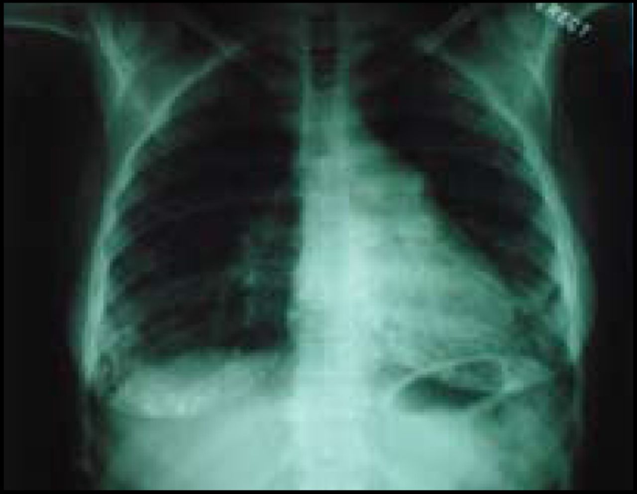
Scleroderma and PAH
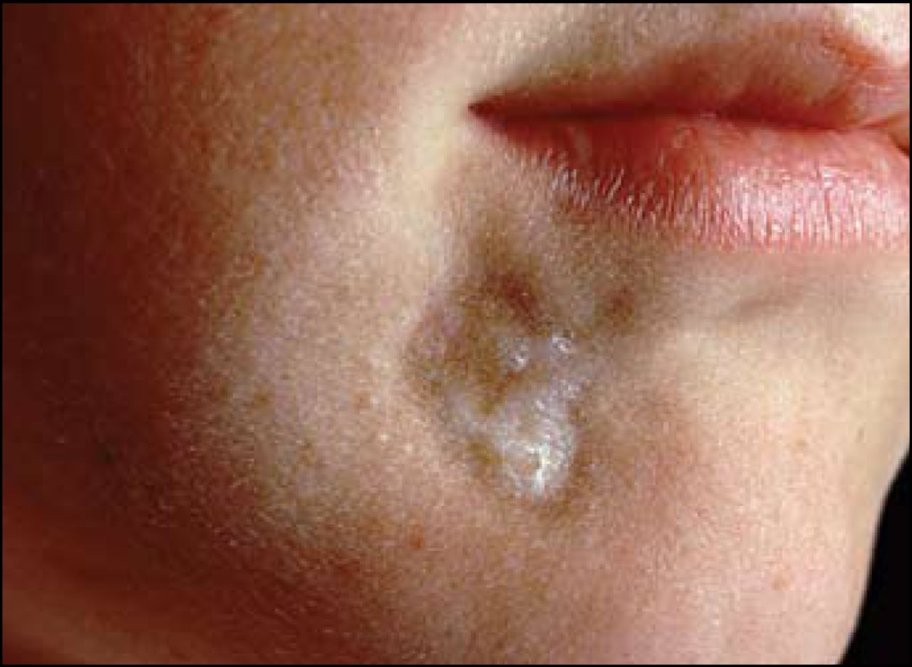
Scleroderma (SSc) affects the connective tissue of the skin, internal organs and the walls of blood vessels. As with SLE, its etiology is unknown and the course and severity of the disease varies. Systemic features may include fibrosis and degeneration of the heart, lungs, kidneys and GI tract. SSc usually affects people between the ages of 30 and 50 years. Women are affected more often than men.
Dr. Virginia Steen, Georgetown University, Washington, DC, stressed the need to assess patients with SSc for the risk of developing pulmonary arterial hypertension (PAH). Early diagnoses and intervention with emerging treatments could prevent more severe PAH and associated heart failure. Patients who develop severe, isolated or vasculopathic PAH often have a long history of Raynaud's disease, limited scleroderma, an anticentromerer antibody, an extremely low diffusing capacity (DLCO), a near normal forced vital capacity (FVC), minimal fibrosis, and a FVC%/DLCO5 ratio >1.8 at the time of diagnosis. Morphometry studies indicate a correlation between intimal proliferation of pulmonary arteries and disease duration. DLCO values that decreased over time to levels below 50% predicted a vasculopathic type of PAH in most patients. Possible predictors of severe PAH include an elevated echo Doppler pressure, exercise-induced pulmonary pressure, or a combination of these findings, however these are not always clear indicators. A right heart catheterization is the gold standard and is necessary to make the diagnosis of PAH in SSc.
Dr. Steen highlighted four risk factors likely to predict future severe and deadly PAH: dyspnea, a very low DLCO value, a high FVC/DLCO ratio, and an increased PASP at rest or exercise. “The treatment of PAH is changing every year, and we have not yet developed overall treatment protocols for scleroderma, but we can work towards improving outcomes of patients with severe lung disease.”
Future Treatments, New Hope
Dr. David Isenberg, University College, London, UK, discussed future treatment directions for SLE. Although the 10-year survival rates for patients with SLE have improved dramatically and are approaching 90%, the potential for significant morbidity and mortality remains. Dr. Isenberg believes that survival rate improvements can be achieved by using a combination of earlier diagnosis at milder disease states, multiple serological tests, use of steroids and other immunosuppressive agents, and the availability of renal dialysis and transplantation. He said “We are moving from an era of a rather random treatment approach with immunosuppressives like azathioprine, cyclophosphamide, or methotrexate to more specific targeting of key molecules known to be essential to the development of SLE.” These newer treatments include mycophenolate mofetil, B cell depletion, biological agents, and haemotopoietic stem cell transplantation (Clin Exp Immunol 2005;140:205–12).
According to Dr. Isenberg, mycophenolate mofetil is a safer substitute for cyclophosphamide and selectively suppresses T and B lymphocyte proliferation. This causes suppression of antibody synthesis and glycosylation of adhesion molecules and cytokine antagonism, which play a role in the pathogenesis of SLE. Though remission rates are similar to cyclophosphamide, there are fewer adverse events.
For SLE patients unresponsive to conventional therapies, B cell depletion therapy with the anti-CD20 monoclonal antibody rituximab is effective. Of five patients treated with rituximab, cyclophosphamide, and high dose oral corticosteroids, two were disease-free for 2 to 3 years post-B cell depletion. Reduction in disease activity, improvements in renal function and immunological and haematoligcal indices were reported in the remaining patients [Arthritis Rheum 2003;48(Suppl9):S924].
Another alternative, immunoblation followed by autologus haematopoietic stem cell transplant (HSCT), has been used successfully in a variety of autoimmune diseases. Although studies have demonstrated that HSCT can induce SLE remission, it appears to be curative in less than 50% of patients and the adverse effect profile, including infection, organ dysfunction, and other long-term toxicities are yet to be defined.
Referencing a number of new biological therapies that are being studied such as anti-TNF-alpha (Ann Rheum Dis 2005;64:403–07), the targeted immunosuppression of CD40 ligand/CD40or CTLA-4/CD28/CD80/CD86 (Lupus 2005;14:197–203), and recombinant DNA, Dr. Isenberg concluded: “The treatment of SLE is entering into a new era that will be all encompassing with pharmacological therapies tailored to the individuals' symptoms and disease.”

- © 2006 MD Conference Express
Tools


Table of contents
Cited By...
- No citing articles found.