

Summary
This article discusses the clinical markers for osteoarthritis (OA), a study looking at the relationship between changes in bone marrow edema (BME) and clinical features in patients with OA, joint distraction treatment for OA, pharmacological approaches to OA treatment, as well as EULAR 2006 recommendations for the management of hand OA, among other things.
- arthritis
Dr. Maxime Dougados, University René Descartes, Paris, France, discussed the clinical markers for osteoarthritis (OA). He believes that the most important symptom is pain (Arthritis Rheum 2004;50:1360–5) with the Lesquene index at baseline being a strong predictor of progression. Suggesting that the source of the pain may be an inflammatory episode of OA synovitis (Ann Rheum Dis 2005;64:1703–09), Dr. Dougados referred to the results of a 2-year prospective follow-up study that indicated that baseline radiographic and subjective symptomatic severity and inflammation were predictive of articular replacement (Presented at ACR 2005. Abstract No.1355).
Changes in cartilage morphology appear to be associated with clinical symptoms and time to knee arthroplasty. According to Dr. Felix Eckstein, Paracelsus Private Medical University, Salzburg, Austria, magnetic resonance imaging (MRI) because of its ability to evaluate the volume, thickness, and structural composition of articular cartilage holds significant promise for use in epidemiologic studies of OA progression and clinical trials of treatment response to disease modifying OA drugs (DMOADs). Quantitative assessment of cartilage morphology (qMRI), as well as appropriate image analysis techniques, display high accuracy and adequate precision (RMS CV% between 1 and 3%) for cross sectional and longitudinal studies in OA patients. Longitudinal studies (at 1.5 T) suggest that changes in cartilage volume of 2% to 5% occur annually in OA in most knee compartments. He sees promise in using compositional methods in expanding analysis to the structural and biochemical composition of the cartilage.
Limitations of traditional OA assessment techniques have led to the development of two clinically significant, highly tissue-specific markers that provide information on cartilage synthesis (immunoassays for N-propeptide of type IIA [PIIANP] and IIB collagens) and degradation (fragments of the helical [Helix-II] or C-telopeptide [CTX-II] portion of type II collagen). According to Dr. Patrick Garnero, INSERM and SYNARC, Lyon, France, because Helix-II and CTX-II reflect distinct enzymatic pathways of type II collagen degradation, a combined measurement is more effective in identifying patients with rapidly progressive hip OA. Stating that other markers might also have a role in predicting disease progression, Dr. Garnero discussed results of two studies he conducted in patients with symptomatic knee OA. In the first study, patients with levels of urinary CTX-II above the upper limit of age-matched controls had a 2–3-fold increased risk of progression after 1 year (Arthritis Rheum 2002;46:2613–24), as assessed by radiography or arthroscopy. The predictive value of urinary CTX-II was later confirmed in larger cohorts of patients with knee or hip OA followed over periods of 3–6 years (Arthritis Rheum 2003;48:S683). Dr. Garnero told the audience that it is likely that a combination of several biochemical markers will be required to adequately predict disease progression because of the complex involvement of bone, cartilage and synovium tissue in OA joint damage.
Dr. Peter R. Kornaat, Leiden University Medical Center, Leiden, Netherlands, presented the results of a prospective study conducted to evaluate the relationship between changes in bone marrow edema (BME) and clinical features in patients with OA. Knee MRIs were obtained at baseline and at 2 years from 182 patients diagnosed with familial symptomatic OA at multiple joint sites (GARP-study). In this study, no association between BME change in size over a two-year time period and WOMAC scores (pain, stiffness and function) was found (p>0.05) leading to the conclusion that in the majority of OA patients BME lesions fluctuate in volume over a 2 year time period and BME on a single MRI does not have any predictive value, in opposition to recent studies published by Felson's group (Ann Intern Med 2003;139:330–36).
Dr. Reuben Gobezie 1, Case Western Reserve University, Cleveland, OH, presented the results of a prospective controlled study that analyzed synovial fluid from the knee using tandem mass spectrometry and bioinformatics analysis to identify a protein biomarker profile for OA. Synovial fluid from 42 age-matched patients with early (n = 21) and late OA (n = 21) and 20 healthy subjects was analyzed. The tandem mass spectrometric analysis identified 342 proteins. A protein biomarker profile for OA with a panel of 15 proteins was identified. The p-value for each of these proteins was <0.0001 and each had a rank order within the top 100 proteins on PCA analysis. This is the first study to present a protein biomarker profile for early and late OA.
According to Dr. Floris LaFeber, University Medical Center Utrecht, Netherlands, joint distraction may become the treatment of choice in relatively young patients (55 to 60 years) with severe end-stage knee OA.
The goal of joint distraction is to relieve mechanical stresses on cartilage by using an external fixation frame that prevents further wear and tear of cartilage and allows the chondrocytes to initiate repair. Results from a prospective study in 17 patients with severe ankle OA conducted by Dr. Lafeber showed that after 2 years of follow-up only 4 patients had poor results that led to arthrodesis within one year. Clinical symptoms improved in 66% of the remaining 13 patients; effects were progressive in the 2nd year of follow-up (Osteoarthritis Cartilage 1999;7:474).
Although current literature on clinical and scientific experience with joint distraction is limited, there is a steady spreading of this technique among clinicians and scientists. Further research and analysis will be necessary to understand, validate, and refine this novel approach.
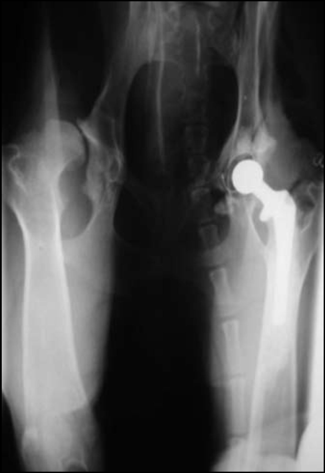
Aseptic prosthesis loosening (APL) is a major problem in orthopedic surgery, said Dr. Thomas Pap, University Hospital Munster, Germany, and it has become increasingly clear that fibroblast-like cells contribute significantly to the initiation and perpetuation of aseptic loosening and the destruction of periprosthetic bone. Dr. Pap presented the results of an animal model that allows for the study of early events in synovial-like interface membrane (SLIM) formation. Results of this study using human SLIM samples and prosthesis loosening fibroblasts (PLF) showed that activated PLF enhance and contribute to bone resorption through superficial erosion of the osseous matrix. According to Dr. Pap, investigating the role of PLF may result in the development of novel and specific therapeutic strategies for APL and other, related conditions.
Dr. Steven Abramson, New York University Hospital for Joint Disease, NY, updated the attendees on pharmacological approaches to OA treatment. He presented three major unsettled questions in OA therapy: the role of the non-pharmaceutical agents; the cardiovascular (CV) safety of traditional NSAIDs vs COX-2 inhibitors; and standards for structure modification. According to Dr. Abramson, findings from RCTs and systematic reviews support a role for glucosamine (GS) and chondroitin sulfate, viscosupplementation, and diacerhein in disease modification with intriguing data showing a positive benefit on delayed time to joint replacement for GS. However, most of the studies have not met regulatory requirements for radiographic outcomes. Dr. Abramson pointed out that there are currently no FDA approved therapies for structure modification in OA. The CV safety of traditional NSAIDs and COX-2s has been much in the news. However, a meta-analyses of RCTs showed no difference among the coxibs vs non-naproxen NSAIDs with respect to CV events and MI (both p=0.3) or vascular death (p=0.09), and a significantly lower incidence of stroke (p=0.03) (BMJ 2006;332:1302–08).
According to Dr. Christiansen, HS Frederiksberg Hospital, Copenhagen, Denmark, “Preliminary results of a systematic review and meta-analysis of three RCTs involving more than 400 patients with knee OA show that weight loss is associated with significant symptom relief.” Clinically relevant improvement in physical disability was seen following a moderate (>5%) weight loss within a 20-week period; a moderate-to-large clinical effect can be achieved with a 10% loss of body weight over a 12-week period. Dr. Christiansen stated that physicians should encourage their overweight OA patients to loose weight.
Only one randomized placebo-controlled double-blind trial of intraarticular (IA) steroids in hip OA has been published, showing a short term symptomatic effect (Osteoarthritis Cartilage 2006;14:163–70). Since placement of IA steroids without imaging guidance is frequently inaccurate, Professor Walter P. Maksymowych, University of Alberta, Edmonton, Canada, and colleagues conducted a RCT to assess the efficacy of image-guided IA. Patients received fluoroscopically guided IA injection and were randomly allocated to placebo (2 ml normal saline) or treatment (40 mg triamcinolone hexacetonide). Primary outcome was improvement in pain two months post-injection determined by the WOMAC scale. Responders were defined as those with ≥ 20% improvement in pain. Subjects receiving IA steroids improved significantly compared with placebo and demonstrated significant gains from baseline in pain, stiffness, physical function and global assessment at 2 months (p<0.001). According to Dr. Maksymowych, this approach should be used in the management of hip OA. The majority of patients will have a response that typically lasts for several months. Injections should be given under fluoroscope guidance.
Dr Michael Doherty, University of Nottingham, UK presented the key EULAR 2006 recommendations for the management of hand OA:
-
Individualized treatment including combined pharmacological and non-pharmacological treatment modalities.
-
Individualized therapy regimens, which take into account: localization of OA; risk factors (age, gender, adverse mechanical factors); type of OA (nodal, erosive, traumatic); presence of inflammation; severity of structural change; level of pain, disability and restriction of quality of life; co-morbidity and co-medication, and patient wishes and expectations.
-
Patients should be educated concerning joint protection and provided with an exercise regime that involves both range of motion and strengthening exercises.
-
Local application of heat, especially prior to exercise, and ultrasound are beneficial treatments.
-
Splints for thumb base OA and orthoses to prevent/correct lateral angulation and flexion deformity are recommended.
-
Local treatments (topical NSAIDs and capsaicin) are preferred over systemic treatments especially for mild to moderate pain and when only a few joints are involved.
-
Paracetamol (up to 4g/day) is the first choice oral analgesic and if successful, preferred for long term treatment.
-
Oral NSAIDs at the lowest effective dose and for the shortest duration should be used in patients who respond inadequately to paracetamol; patients should be re-evaluated periodically. In patients with increased GI risk, non-selective NSAIDs + a gastroprotective agent, or selective COX-2 inhibitor should be used. In patients with increased CV risk, coxibs are contraindicated; non-selective NSAIDs should be used with caution.
-
Symptomatic Slow-Acting Drugs in Osteoarthritis (SYSADOA; e.g., glucosamine, chondroitin sulphate, diacerhein) may give symptomatic benefit with low toxicity, but effect sizes are small, suitable patients are not defined, and clinically relevant structure modification and pharmacoeconomic benefits have not been established.
-
IA injection of long-acting corticosteroid is effective for painful flares of OA, especially trapezometacarpal joint OA.
-
Surgery is an effective treatment for severe thumb base OA and should be considered in patients with marked pain and/or disability when conservative treatments have failed.
- © 2006 MD Conference Express
Tools


Table of contents
Cited By...
- No citing articles found.