

Summary
This article presents data from a study that was conducted to analyze in vivo human anterior cruciate ligament (ACL) graft motion during activities of daily living in patients who had received bone—patellar tendon—bone (BTB) or hamstring (HS) autografts. The results showed that BTB autografts have more femoral tunnel motion than HS autografts 6?weeks after ACL reconstruction.
- Orthopaedic Procedures
- Hip & Knee Conditions
- Sports Medicine Clinical Trials
- Orthopaedic Procedures
- Hip & Knee Conditions
- Sports Medicine
- Orthopaedics Clinical Trials
- Orthopaedics
James N. Irvine, Jr, MD, University of Pittsburgh, Pittsburgh, Pennsylvania, USA, presented data from a study that was conducted to analyze in vivo human anterior cruciate ligament (ACL) graft motion during activities of daily living in patients who had received bone—patellar tendon—bone (BTB) or hamstring (HS) autografts. The results showed that BTB autografts have more femoral tunnel motion than HS autografts 6 weeks after ACL reconstruction.
Graft type is one of numerous surgical variables that influences graft-tunnel healing following ACL reconstruction. However, the optimal choice of graft remains controversial because of a lack of evidence-based consensus available to guide surgeons' decision making. According to Dr. Irvine, although clinical and kinematic outcomes of BTB and HS autografts are similar, data from animal studies have suggested that their healing processes may differ.
With this in mind, Dr. Irvine and colleagues conducted a prospective study to compare postoperative BTB and HS graft motion within the femoral and tibial tunnels and the intra-articular graft. They hypothesized that BTB autografts would have less intraosseous tunnel motion, greater midsubstance strain, and less anterior tibial translation than HS autografts at 6 weeks following ACL reconstruction as a result of faster osteointegration.
The study included 12 patients (ages 16 to 37 years; mean age, 24 years) who received BTB (n = 6) or HS (n = 6) autografts. A single surgeon performed anatomic single-bundle ACL reconstruction through a medial portal with the same technique for ACL tunnel placement and used suspensory fixation in all cases. Outcome measurements included graft motion within bone tunnels, midsubstance ACL strain, and knee kinematics at 6 weeks postoperatively.
Prior to implantation, six .8-mm tantalum beads were embedded into each graft, pairs of which were located within each bone tunnel and in the graft midsubstance. Computed tomographic scans were obtained 6 weeks postoperatively and used to create 3-dimensional femur and tibia bone models. Cylindrical coordinate systems were then fit to the bone tunnels to examine tunnel motion, and dynamic stereo x-ray images were collected while patients walked on a treadmill and descended stairs. Longitudinal (along tunnel axis) and transverse (within tunnel cross section) graft motion was quantified by 2 methods. One was described as graft excursion, defined as the total path distance traveled by the graft over a set period of time (walking: 200 ms following foot strike; stair descent: 300 ms following single-leg support).
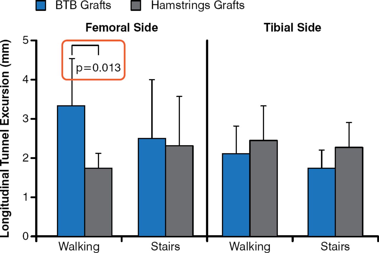
Postoperative rehabilitation was similar in both groups, with all patients having returned to their activities of daily living at 6 weeks postoperatively. The grafts were still moving in all patients within the femoral and tibial bone tunnels at the 6-week follow-up testing. The BTB group displayed significantly more longitudinal graft excursion within the femoral tunnel compared with the HS patients (Figure 1). It is important to note that all patients were doing well at follow-up. The findings of this study showed no evidence of faster graft osteointegration of BTB over HS, no detectable midsubstance strain because the grafts were still moving within the bone tunnels, and no difference in knee kinematics between the grafts, Dr. Irvine said.

Longitudinal Graft Motion in Bone Tunnels 6 Weeks After ACL Reconstruction
ACL, anterior cruciate ligament; BTB, bone—patellar tendon—bone autograft.
Reproduced with permission from J. N. Irvine, Jr, MD.
On November 12, 2014, the bracket was added to this figure.
Dr. Irvine acknowledged that some limitations of this study included its small sample size and the absence of contralateral knee data. Although 1-year follow-up testing remains under way for this trial to assess whether the pattern of findings in this study changes as healing progresses, future studies using quantitative magnetic resonance imaging will be essential to further assess graft healing.
- © 2014 MD Conference Express®
Tools


Table of contents
Cited By...
- No citing articles found.