

Summary
Atherosclerosis is recognized as a complex inflammatory condition in which various immune system cells interact with each other and with cells of the arterial wall. This article describes inflammation as the underlying mechanism that links obesity and atherosclerosis.
- Obesity
- Cardiometabolic Disorder
- Inflammatory Disease
- Hypertensive Disease

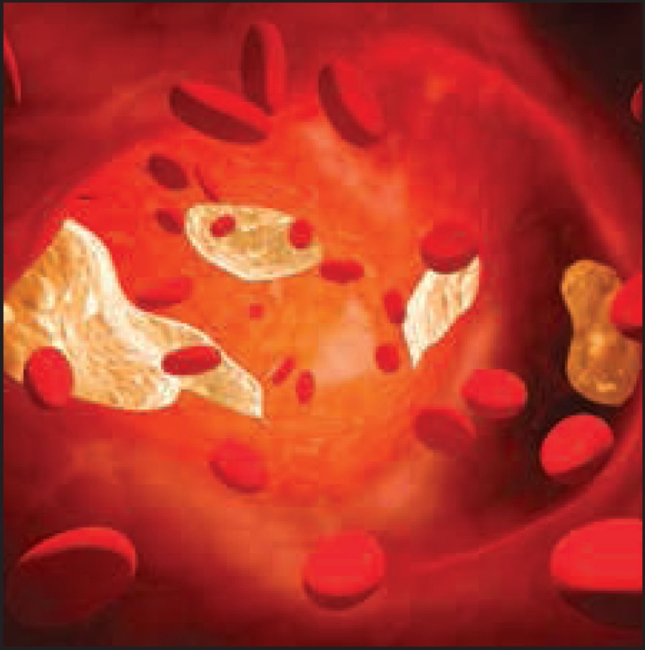
Just a few decades ago, atherosclerosis was viewed as a lipid storage disease, in which cholesterol accumulated as a waxy deposit within the walls of an inanimate artery, similar to rust clogging a pipe. Today, atherosclerosis is recognized as a complex inflammatory condition in which various immune system cells interact with each other and with cells of the arterial wall. Peter Libby, MD, Harvard Medical School, Boston, Massachusetts, USA, described inflammation as the underlying mechanism that links obesity and atherosclerosis.
Natural History of Atherosclerosis
Within normal blood vessels in homeostasis, inflammatory cells are not typically present at the vessel wall. But when the vessel endothelium is exposed to atherogenic stimuli, such as dyslipidemia, diabetes, and hypertension, endothelial cells express adhesion molecules that attract leukocytes. In response to chemoattractant messages, leukocytes adhere to the vessel wall and enter the arterial intima. Monocytes are recruited in the largest number among all leukocytes. In the presence of hyperlipidemia, monocytes propagate the innate immune response by expressing proinflammatory cytokines and differentiating into proinflammatory macrophages.
Monocyte-derived macrophages absorb modified lipoproteins to become foam cells, the characteristic cells of the initial atherosclerotic lesion [Libby P et al. Circulation 2007]. Other cells of the innate immune response, including mast cells, also are recruited through proinflammatory signaling to participate in atherogenesis.
Innate Immunity and Atherosclerosis
Adipose tissue is a source of cytokines that mediate systemic inflammation, insulin resistance, and endothelial dysfunction, all of which promote atherosclerosis. Adipose tissue macrophages mediate some of the inflammatory changes that are characteristic of obesity. Two distinct populations of macrophages have been identified within adipose tissue. Proinflammatory macrophages, which are more predominant in visceral adipose tissue, secrete inflammatory cytokines, such as tumor necrosis factor (TNF) and inducible nitric oxide synthase (iNOS). Anti-inflammatory macrophages secrete anti-inflammatory cytokines that quell inflammation and facilitate tissue repair [Rocha VZ et al. Nat Rev Cardiol 2009].
Imaging studies have been instrumental in demonstrating the metabolic differences between visceral and subcutaneous adipose tissue. As body weight increases, proinflammatory macrophages accumulate in visceral adipose tissue. In a study of adipose tissue in mice that consumed a high-fat diet, obesity that was associated with increased expression of TNF and iNOS rose, indicating enhanced activity of proinflammatory macrophages [Lumeng CN et al. J Clin Invest 2007]. Using whole-body fluorodeoxyglucose positron emission tomography (FDG-PET) scans, Dr. Libby and colleagues showed that glucose uptake is significantly greater in macrophage-rich visceral adipose tissue compared with subcutaneous adipose tissue [Christen T et al. J Am Coll Cardiol Img 2010].
Adaptive Immunity and Atherosclerosis
Obesity is associated with increased accumulation of T cells in adipose tissue. Recent research suggests that T cells play an important role in modulating the immune response to atherogenesis. T cells enter arterial plaque by interacting with adhesion molecules on the surface of endothelial cells, in response to chemoattractants, such as regulated on activation, normal T cell expressed and secreted (RANTES) [Wu H et al. Circulation 2007].
Once in the vessel wall, type 1 helper T (Th1) cells can induce proinflammatory responses in macrophages by releasing interferon (IFN)-gamma [Nishimura S et al. Nat Med 2009; Rocha VZ et al. Circ Res 2008]. Another population of regulatory T cells, type 2 helper T (Th2) cells, produces interleukin (IL)-4 and IL-13. These cytokines attenuate many of the highly proinflammatory effects of IFN-gamma. Therefore, through the proinflammatory effects of IFN-gamma and the anti-inflammatory effects of IL-4 and IL-13, T lymphocytes regulate signals that promote the accumulation of macrophages within visceral adipose tissue [Rocha VZ et al. Nat Rev Cardiol 2009].
Clinical Implications and New Therapeutic Opportunities
Clinicians now understand that obesity, inflammation, and atherosclerosis are closely linked. Visceral fat accumulation contributes to the systemic inflammation that drives atherosclerosis and its complications, including arterial stenosis and thrombosis. Within atherosclerotic plaques, increased activation of inflammatory cells promotes thrombosis and increases the risk of acute coronary syndromes. Recognizing these pathways opens up new possibilities for therapy, including a new focus on anti-inflammatory therapies for the management of atherosclerosis.
New options for risk assessment are also available. For instance, biomarkers, such as high-sensitivity C-reactive protein (hsCRP), are emerging as important markers of systemic inflammation. In patients with diabetes, elevated levels of plasminogen activator inhibitor type 1 (PAI-1) in blood and in the arterial wall may favor decreased fibrinolysis and accelerated atherosclerosis [Calles-Escandon J et al. Diabetes 1998; Pandolfi A et al. Arterioscler Thromb Vasc Biol 2001]. After adjusting for body mass index (BMI), hsCRP correlates with abdominal visceral adiposity. In addition, hsCRP levels predict cardiovascular events in apparently healthy persons and in those with manifest atherosclerotic disease, independently of traditional risk factors. Inflammatory biomarkers can guide the use of statins and perhaps other therapies that modify systemic inflammation in patients with atherosclerosis.
- © 2011 MD Conference Express
Tools


Table of contents
Cited By...
- No citing articles found.