

Article Figures & Data
Figures
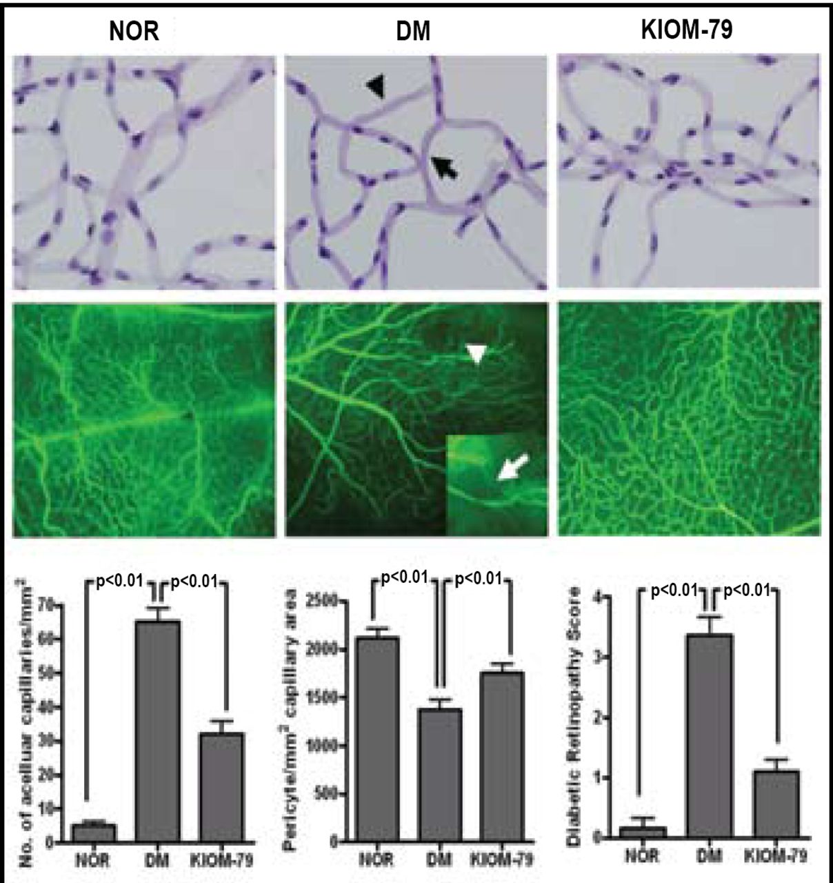
- Figure 1.
Effects of KIOM-79 on Retinal Vascular Damage.
Top panel: Immunohistochemistry of retinal samples showing pericyte ghost (arrow) and acellular capillary (arrowhead).
Middle panel: Fluorescein angiography showing nonperfusion areas and vessel-narrowing (magnified inset).
Bottom panel: Quantitative analysis of acellular formation, pericyte ghosts, and retinopathy score of retinal vessels. Nor = normal, untreated rat; DM = ZDF rat treated with placebo; KIOM-79 = ZDF rat treated with KIOM-79.

Tools
KIOM-79 Slows the Development of Diabetic Retinopathy in Animal Models
Permalink:


Table of contents
Cited By...
- No citing articles found.