

Summary
Good quality images are determined by number of dimensions, spatial resolution, signal-to-noise ratio, image contrast, and the presence of artifacts. This article presents an overview of 3D image acquisition with computed tomography and magnetic resonance from the perspective of obtaining accurate images suitable for 3D printing. Other topics include a review of the art of 3D visualization vs 3D printing, as well as the role of rapid prototyping using 3D printing technologies to assist surgeons in optimizing surgical decisions.
- imaging
- three dimensional
Karin E. Dill, MD, University of Chicago, Chicago, Illinois, USA, presented an overview of 3D image acquisition with computed tomography (CT) and magnetic resonance (MR) from the perspective of obtaining accurate images suitable for 3D printing. Good quality images are determined by number of dimensions, spatial resolution, signal-to-noise ratio, image contrast, and the presence of artifacts.
Typical MR slices are 1 to 10 mm thick and CT slices range from 0.5 to 5 mm. The anatomic structure may not be perfectly homogeneous across each slice. Two-dimensional imaging, therefore, should be considered an average of the image information. Two-dimensional images are representative of tissue slices, while 3D images reflect a volume rather than a slice.
During CT, cross sections of the body are irradiated. CT generates 2D radiographic images taken around a single axis of rotation, obtained in an arc in the transverse plane. They provide one channel of spatial data.
Three-dimensional images can be generated in the axial plane, perpendicular to the z-axis plane. Multisection CT scans with multiple rows of detectors along the longitudinal (z) axis of the patients can generate stacks of data for 3D images. Further, 3D imaging offers a more complete representation of all the tissue in a given region, the ability to postprocess and reformat images into any slice plane, and a better representation of tissue geometry and relationships between structures.
Spatial resolution is related to the ratio of field of view and number of voxels in the image or matrix. Voxels that are the same size in all 3 dimensions (x, y, z) are referred to as isotropic voxels. Spatial resolution defines the smallest features that may be detected in the image. This can be complicated by the signal-to-noise ratio, which determines the ratio of the useful data to the nonuseful data, and must be adequate for tissue signals in routine CT and MR scans.
Michael L. Steigner, MD, Brigham and Women's Hospital and Harvard Medical School, Boston, Massachusetts, USA, reviewed the art of 3D visualization vs 3D printing.
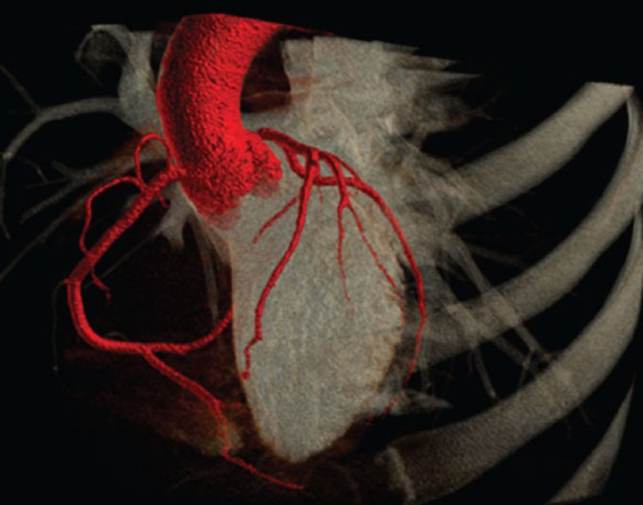
Until recently, heart imaging focused mainly on retrospectively gated computed tomography angiography (CTA). A new technique, prospective gating, has gained attention as a way to reduce radiation dose in addition to improving image quality. With prospective gating, CT data acquisition is synchronized with the cardiac cycle. Images reconstructed during the cardiac phases with minimum motion are of higher quality and have less motion artifact (Figure 1). For instance, a study that assessed the relationship between the phase window width and image quality in prospectively electrocardiogram-gated 320-detector row coronary CTA showed that a phase window width of 10% reduces patient radiation and yields diagnostic images in > 90% of patients [Steigner ML et al. Int J Cardiovasc Imaging. 2009]. Further, heart rate control was found to be an important component of 320-detector row prospectively gated CT dose reduction.

Three-Dimensional Volume Rendered on Multiple-Detector Computed Tomography
Reproduced with permission from ML Steigner, MD.
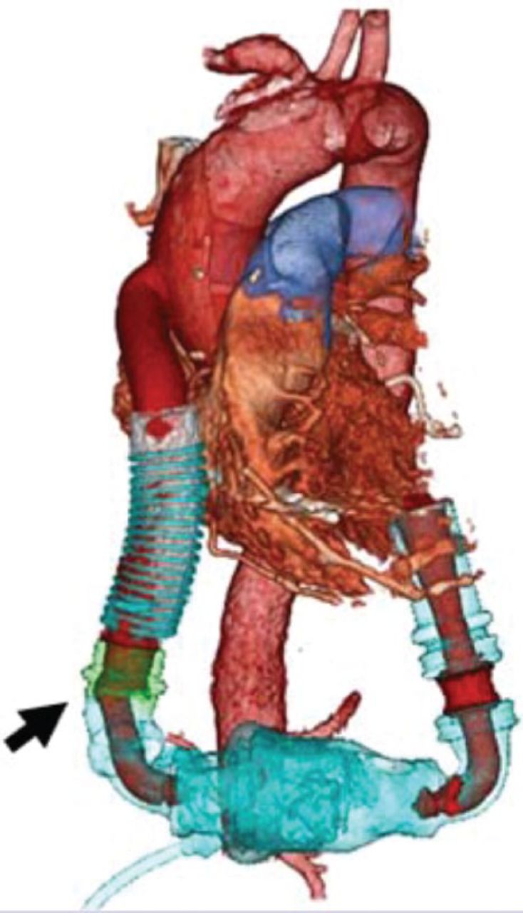
Color correction of the scan can be performed using the Color Look Up Table, which renders objects that may normally be hidden. Dr Steigner described the use of 3D visualization for detecting bend relief disconnection in patients implanted with left ventricular assist devices (Figure 2) [Waller AH et al. Circ Cardiovasc Imaging. 2014].

Three-Dimensional Visualization of Bend Relief Disconnection of Implanted Left Ventricular Assist Device
Arrow demonstrates a complete disconnection of the bend relief from the sealed outflow graft.
Reproduced from Waller AH et al. Evaluation of bend relief disconnection in patients supported by a HeartMate II left ventricular assist device. Circ Cardiovasc Imaging. 2014;7(5):844–848. With permission from American Heart Association, Inc.
Once a good image that reflects the anatomic structure in question has been captured, postprocessing software is used to render the image into a 3D object. The 3D data set can be reformatted using several types of postprocessing algorithms. This might include volume rendering, multiplanar reformation, thin-slab maximum intensity projection, curved multiplanar reformation, angiographic view, and plaque-loaded angiographic view. The interpreter should know the advantage and disadvantage of each postprocessing 2D and 3D image and renderings before the final printing. Multidetector CT is a first-line modality for pretreatment planning and imaging to detect hidden deformities. Proper data collection, surface rendering, and stereolithographic editing can produce detailed skeletal and soft tissue structures from CT and MRI data that cannot be adequately conveyed through a computer screen.
Shi-Joon Yoo, MD, University of Toronto, Toronto, Ontario, Canada, discussed the role of rapid prototyping using 3D printing technologies to assist surgeons in optimizing surgical decisions. Physical heart replicas can fill the gap between the imagination of the medical images and reality, as well as simulate the intended procedures to improve accuracy and reduce procedure and anesthesia time.
Rapid prototyping includes multiple technologies, methods, and materials. Three-dimensional technologies such as selective laser sintering, electron beam melting, inkjet technology, stereolithography (STL), fused deposition modeling, laminated object manufacturing, and 3D microfabrication are routinely being employed for this process.
Once the imaging is complete, the multislice digital images are partitioned into multiple segments that locate objects and boundaries, creating a set of contours that defines the structure. Image segmentation is then used to create a 3D reconstruction using a Digital Imaging and Communications in Medicine (DICOM) data set. DICOM supports the distribution and viewing of medical images from CT, MRI, and other medical modalities. The DICOM data are converted STL for manufacturing with resin and solidification with UV light.
Rapid prototyping of vessel and heart deformities as 3D replicas is feasible and can aid in planning procedural time, dosing, and cost. In congenital heart disease surgery, the replicas are of tremendous help for making surgical decisions.
- © 2015 MD Conference Express®
Tools


Table of contents
Cited By...
- No citing articles found.