

Article Figures & Data
Figures
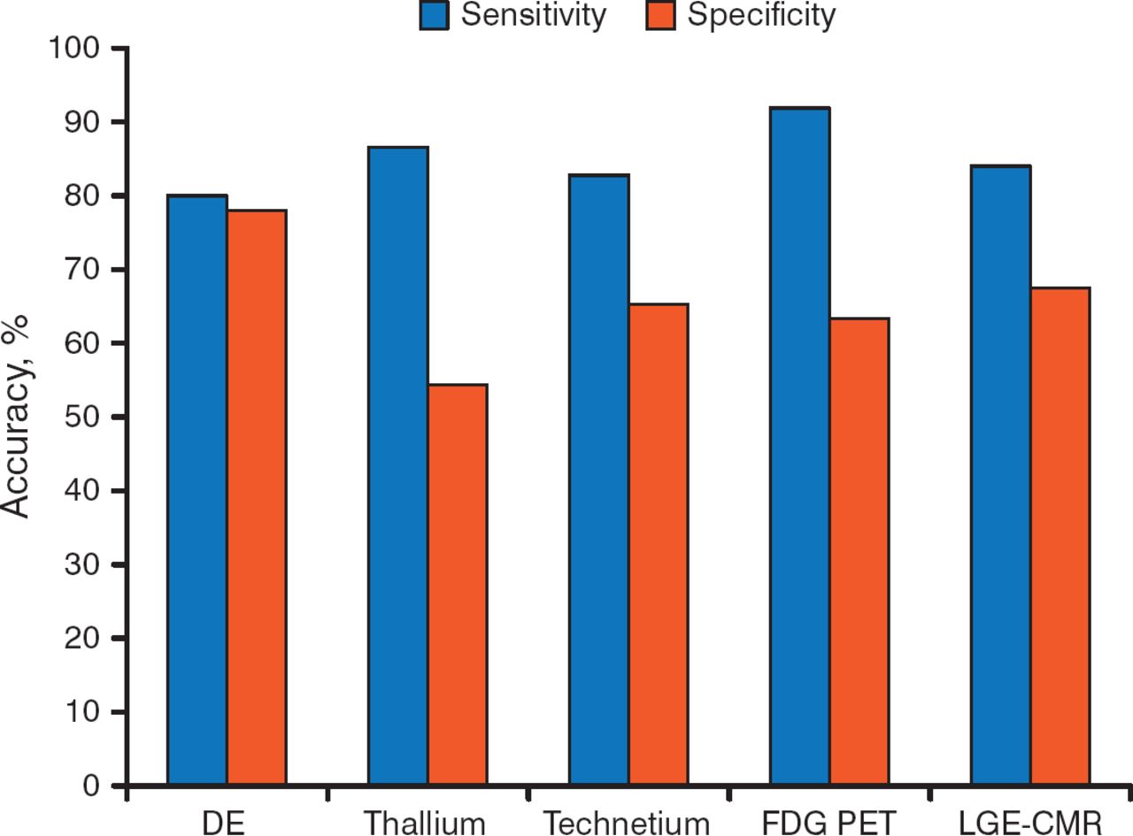
- Figure 1.
Sensitivity and Specificity of Various Imaging Methods for Identifying Myocardial Viability
DE, dobutamine echocardiography; FDG PET, F-18 fluorodeoxyglucose positron-emission tomography; LGE-CMR, late gadolinium enhancement cardiovascular magnetic resonance.
Source: Schinkel AF et al. Curr Probl Cardiol. 2007. Reproduced with permission from A Fathala, MD.
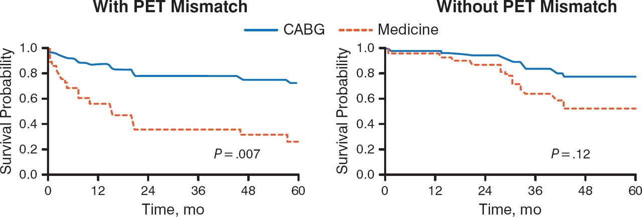
- Figure 2.
Prognosis of Patients With LV Dysfunction by PET Pattern of Viability and Mode of Treatment
CABG, coronary artery bypass grafting; LV, left ventricular; PET, positron-emission tomography.
Adapted from the Journal of Thoracic and Cardiovascular Surgery, 116, Di Carli MF et al, Long-term survival of patients with coronary artery disease and left ventricular dysfunction: Implications for the role of myocardial viability assessment in management decisions, 997–1004, Copyright (1998), with permission from Mosby, Inc.
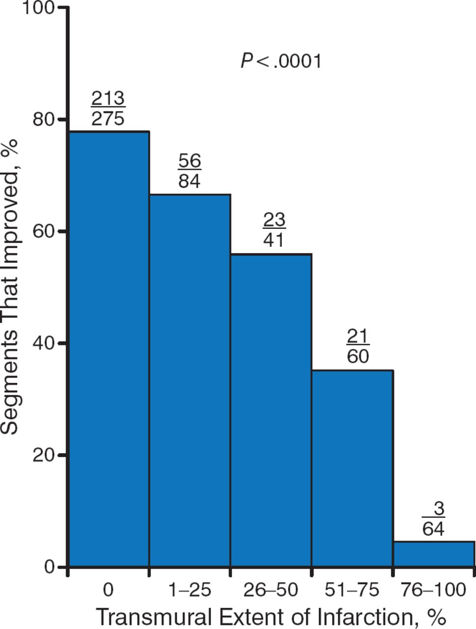
- Figure 3.
LGE Identifies Reversible Myocardial Dysfunction Prior to Revascularization
LGE, late gadolinium enhancement.
Source: Kim RJ et al. N Engl J Med. 2000. Reproduced with permission from A Fathala, MD.
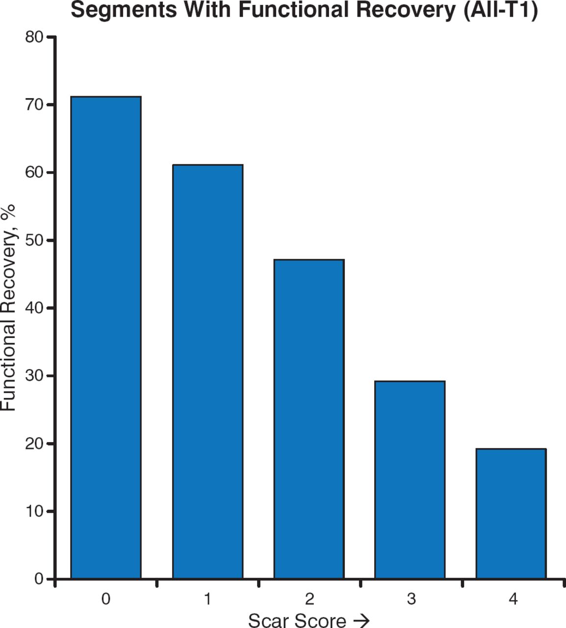
- Figure 4.
Inverse Relationship Between Extent of Scar and Functional Recovery
Adapted from Lenge VV et al. 124 Delayed-enhancement MRI as a predictor of functional recovery after revascularization: results from an International Multicenter Viability Trial. J Cardiovasc Mag Res. 2008;10:A25. With permission from Lenge VV et al and BioMed Central.


Tools
Imaging Solutions to Determining Myocardial Viability before Revascularization
Permalink:


Table of contents
Cited By...
- No citing articles found.