

Article Figures & Data
Figures
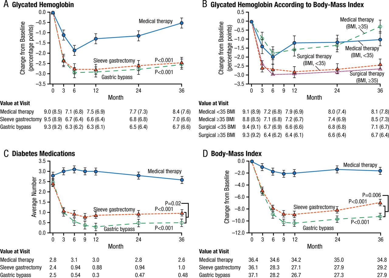
- Figure 1.
Mean Changes in Parameters of Diabetes Control
Mean Changes in Measures of Diabetes Control from Baseline to 3 Years. Shown are the percentage change in glycated hemoglobin levels (Panel A), the percentage change in glycated hemoglobin levels according to body-mass index (BMI) (Panel B), the average number of diabetes medications during the study period (Panel C), and the changes in BMI (Panel D) over a 3-year period among patients receiving intensive medical therapy only, sleeve gastrectomy, or gastric bypass. I bars indicate standard errors. Mean values in each group are provided below the graphs; in Panels A and B, median values are also provided in parentheses. P values are for the comparison between each surgical group and the medical-therapy group in Panels A, C, and D. In Panel B, P=0.008 for the comparison between the surgical groups and the medical-therapy group for the subgroup of patients with a BMI of less than 35; P<0.001 for the comparison for the subgroup with a BMI of 35 or more.
From N Engl J Med, Schauer PR et al., Bariatric Surgery versus Intensive Medical Therapy for Diabetes—3-Year Outcomes, Volume No. 370, Page No. 2002-2013. Copyright © (2014) Massachusetts Medical Society. Reprinted with permission from Massachusetts Medical Society.
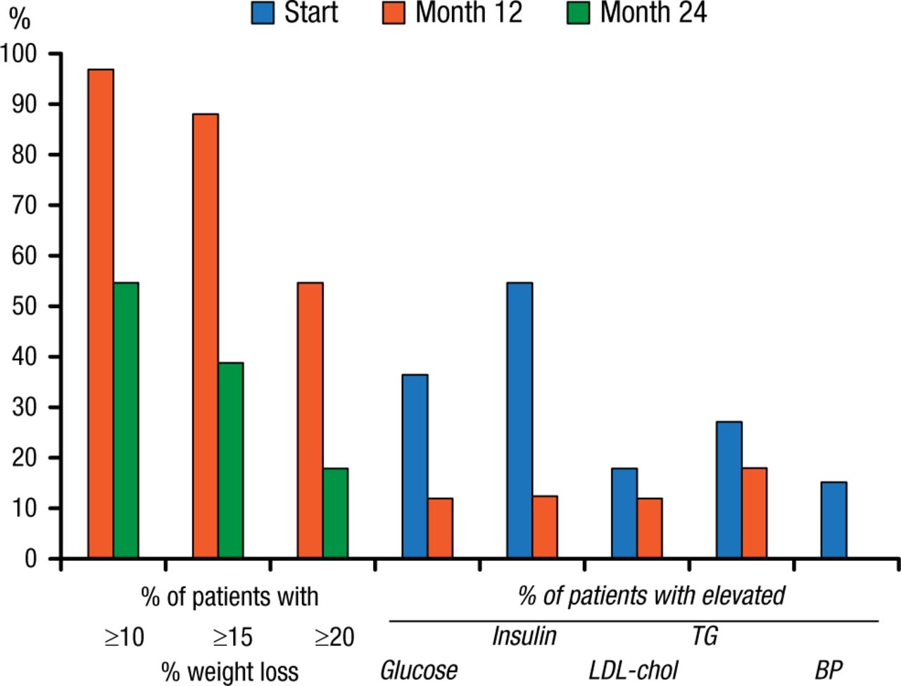
- Figure 2.
Weight and Comorbidity Reductions Following Intragastric Balloon Therapy
Per protocol analysis. Percentage of patients achieving 10%, 15%, and 20% weight loss after 12 and 24 mo, and percentage of patients with elevated values for glucose (> 6 mmol/L), insulin (>20 IU/L), LDL-cholesterol (>4.5 mmol/L), triglycerides (TG) (>2 mmol/L), and elevated diastolic blood pressure (BP) (>100 mm Hg) at baseline (start) and after 1 year.
Reprinted from Gastrointest Endosc, Vol 61, Mathus-Vliegen EMH et al., Intragastric balloon for treatment-resistant obesity: safety, tolerance, and efficacy of 1-year balloon treatment followed by a 1-year balloon-free follow-up, Pages 19-27, Copyright (2005), with permission from American Society for Gastrointestinal Endoscopy.


Tools


Table of contents
Cited By...
- No citing articles found.