

Summary
Radiomics is opening new possibilities for data collection and improved decision making in treating cancer. This article addresses the advances in the use of radiomics and radiogenomics and how this will affect the future practice of radiologists.
- tomography
- oncology genomics
Radiomics is opening new possibilities for data collection and improved decision making in treating cancer. In this session, the plenary speakers addressed the advances in the use of radiomics and radiogenomics and how this will affect the future practice of radiologists.
RADIOMICS AND DATA COLLECTION FOR DECISION MAKING
Robert J. Gillies, PhD, Moffitt Cancer Center, Tampa, Florida, USA, addressed ways in which data can be collected, curated, and mined to aid in research and clinical decision making. While there have been recent advances in lowering cancer death rates, these are largely attributable to preventive and screening measures. Even new well-targeted medications only can prolong lives, not provide a cure, as Dr Gillies illustrated with the examples of non-small cell lung cancer with EGFR mutations [Sequist LV et al. J Clin Oncol. 2008] and HER2-positive breast cancer [Vogel CL et al. J Clin Oncol. 2002]. These therapies do not cure cancer because tumors are heterogeneous and resistant populations of cells emerge. Because of this heterogeneity, a biopsy may sample more or less resistant cells and not give a full picture of the cancer [Gerlingler M et al. N Engl J Med. 2012]. This can result in errors in determining treatment.
Heterogeneity within tumors is associated with resistance and arises from genome instability in a highly selective microenvironment [Gillies et al. Nat Rev Cancer. 2012]. By understanding and analyzing these microenvironments using radiologic images (radiomics), it may be possible to quantify this heterogeneity [Gatenby RA et al. Radiology. 2013]. Quantitative features analyzed in this way may be prognostic, as has been demonstrated in some recent publications [O'Connor JP et al. Clin Cancer Res. 2015; Cunliffe AR et al. Phys Med Biol. 2014].
In the future, radiologists will have an expanded role in curating, extracting, and analyzing data. In radiomics, images are converted into data that can be mined and analyzed to relate features to outcomes. These data can also be correlated with genomic data (radiogenomics). Dr Gillies stated that > 600 quantitative features can be extracted from computed tomography (CT) images, including shape-based features (eg, sphericity and compactness) and texture-based features (eg, skewness, kurtosis, and entropy). Features can be grouped into different classes. Agnostic features are described with texture (not words), while semantic features are described with words (eg, size and shape). Habitat features include combinations of features describing specific microenvironments.
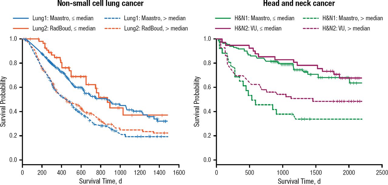
Grove and colleagues published a study showing that the greater the difference in entropy values in different parts of a tumor, the worse the outcomes; additionally, more solid tumors have better prognoses [Grove O et al. PLoS One. 2015]. This information is mildly correlated to stage and histopathology; in other words, it provides additional information or adds value. When the most stable features are used to generate predictions, the same features have value for different cancers (Figure 1) [Aerts HJ et al. Nat Commun. 2014]. Different habitats select for different genotypes, so habitat features may be associated with genotypic heterogeneity [Zhou M. Transl Oncol. 2014].

Survival and Kaplan–Meier Radiomics Signature Based on 100 Most Stable Features
Adapted by permission from Macmillan Publishers Ltd: Nature Communications. Aerts HJWL et al. Decoding tumour phenotype by noninvasive imaging using a quantitative radiomics approach. 2014;5:4006. Copyright (2014).
Large quantities of data are needed to build models, but there are difficulties in using retrospective data. Ideally, information needs to be collected prospectively, put into predictive models, and then put into longitudinal models to assess the effectiveness of the models. Serum data, laboratory data, molecular data, and radiomics data should all be included. Because these data have diagnostic, prognostic, monitoring, and predictive value, Dr Gillies noted that it will be important to develop ways to gather and analyze data in efficient ways and with redesigned approaches to image reading, data collection, and decision support [Gillies RJ et al. Nat Rev Cancer. 2012].
RADIOMICS AS A PATHWAY TO PRECISION MEDICINE
Hedvig Hricak, MD, PhD, Memorial Sloan Kettering Cancer Center, New York, New York, USA, discussed ways in which advances in genomics and bioinformatics will shape the future of oncology and oncologic imaging. Precision medicine involves carefully selecting the right treatment at the right time for each patient and requires robust, validated biomarkers [Rubin EH et al. Clin Cancer Res. 2014]; the molecular heterogeneity of cancer poses a tremendous challenge to the implementation of precision medicine and also provides opportunities to use imaging for in vivo diagnostics. The development of radiomics, radiogenomics, and targeted molecular imaging will help to identify differences in molecular characteristics both within and between tumors and supply a wealth of imaging biomarkers. In turn, these imagine biomarkers will help to address some of the most critical biological questions involved in cancer care, including which lesions to biopsy [Vogelstein B et al. Science. 2013].
Radiomics, radiogenomics, and targeted molecular imaging form the next generation of imaging. Because of the tremendous complexity involved in developing these tools, great care must be taken to use appropriate methodologies and lessons can be learned from previous work in omics. It is important to have transparency, a discovery phase, a test validation phase, and an evaluation of clinical utility [Micheel CM et al., eds. Evolution of Translational Omics: Lessons Learned and the Path Forward. Washington, DC: National Academies Press. 2012].
Radiogenomics includes two different areas—radiation therapy and imaging. In radiation therapy, the goal is to identify mutations that put patients at greater risk of radiation toxicity. In imaging, the goal is to look for associations between imaging features and both genetic and epigenetic features. Both epigenetics (heritable changes not in the genetic code) and genetics are involved and influence each other in cancer development [Herceg Z et al. Carcinogenesis. 2013].
Radiomics can provide complementary information to improve diagnosis and treatment selection while also improving dissemination of knowledge, opportunities for education, and tumor characterization. Additionally, it can lead to more precise biopsies.
Dr Hricak described the difficulty in using standard approaches to distinguish between leiomyosarcoma (a rare malignancy) and atypical leiomyoma (cancers that require very different treatments) as shown in the results of one analysis. While highly experienced radiologists outperformed machine analysis, machine learning and technical analysis successfully differentiated between the two (area under the curve 87%). Dr Hricak also gave an example of a prostate cancer tumor for which texture analysis provided improved visualization and characterization, noting that these techniques will help radiologists to more effectively do their work.
Ultimately, the tools of radiomics and radiogenomics could help all radiologists extract larger amounts of clinically relevant information from features seen in imaging and molecular markers. Pilot studies in radiogenomics have looked at candidate genes and gene expression patterns [Karlo CA et al. Radiology. 2013; Gevaert O et al. Radiology. 2012]. One study showed associations between certain CT parameters and clear-cell renal-cell carcinoma mutations, allowing radiologists to recognize and put emphasis on factors (such as renal vein invasion) that are bad prognostic factors [Karlo CA et al. Radiology. 2014]. With the ability to recognize associations between imaging features and genome clusters with prognostic significance, radiologists will be able to recommend testing for relevant genes. In addition, they will be better able to answer questions as to where to biopsy, whether treatment is needed, and how aggressive any treatment needs to be.
Integrated diagnostics refers to a convergence of imaging, laboratory medicine, and molecular pathology to refine assessments. Systems pathology is moving in a similar direction, combining many types of information, and working together is necessary to make progress.
Dr Hricak concluded by emphasizing that interdisciplinary collaboration, teamwork in big data analysis, and new approaches in imaging bioinformatics are needed to fully develop the potential of radiomics.
- © 2015 MD Conference Express®
Tools


Table of contents
Cited By...
- No citing articles found.